The Struggles of Traditional Microscopy
For years, scientists and researchers in fields like pathology and research have relied on traditional microscopes for their work. While these tools provide high precision, they come with challenges that slow down workflows and create discomfort for users.
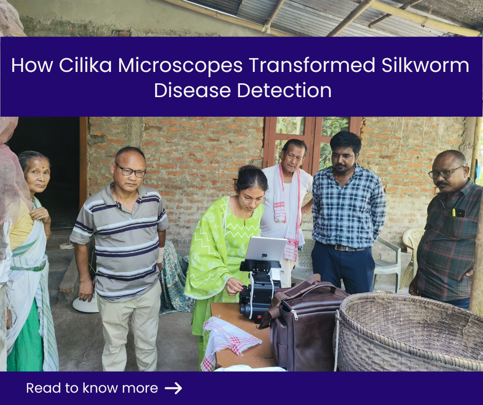
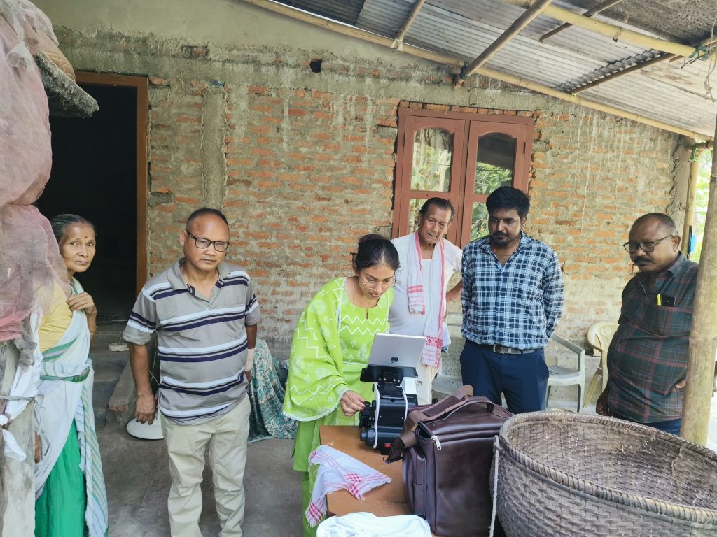
Dr. Arun Kumar K. P., a scientist at Central Muga Eri Research and Training Institute (CMER & TI), Central Silk Board, Ministry of Textiles, Lahdoigarh, Jorhat-Assam, faced these very challenges in his work with silkworm screening. His research required him to analyze thousands of silkworm eggs to detect diseases like Pebrine, a fungal infection that can devastate silk production. However, traditional microscopy made this process slow, exhausting, and inefficient.
The Challenges Faced by Dr. Arun
Dr. Arun’s daily routine involved spending long hours hunched over a conventional microscope, peering through an eyepiece to examine delicate samples. The work was not just tedious—it was physically straining and mentally exhausting. He often experienced neck and back pain due to the constant bending posture, and his eyes would feel fatigued after hours of intense focus. Moreover, the process itself was time-consuming. Adjusting samples manually, documenting observations, and ensuring precision took up most of his day, leaving little room for efficiency.
Another major limitation was the lack of collaboration. Since traditional microscopes allow only one person to view the specimen at a time, sharing insights with colleagues or training students became difficult. Additionally, conducting screenings in the field was almost impossible, as conventional microscopes required a controlled laboratory environment. The lack of portability and adaptability meant that routine diagnosis was often restricted to specific lab conditions, limiting efficiency.
Dr. Arun knew that if he wanted to improve his efficiency and make a real impact on silk production, he needed a better solution. He was searching for a microscope that could enhance accuracy, reduce strain, and make fieldwork possible.
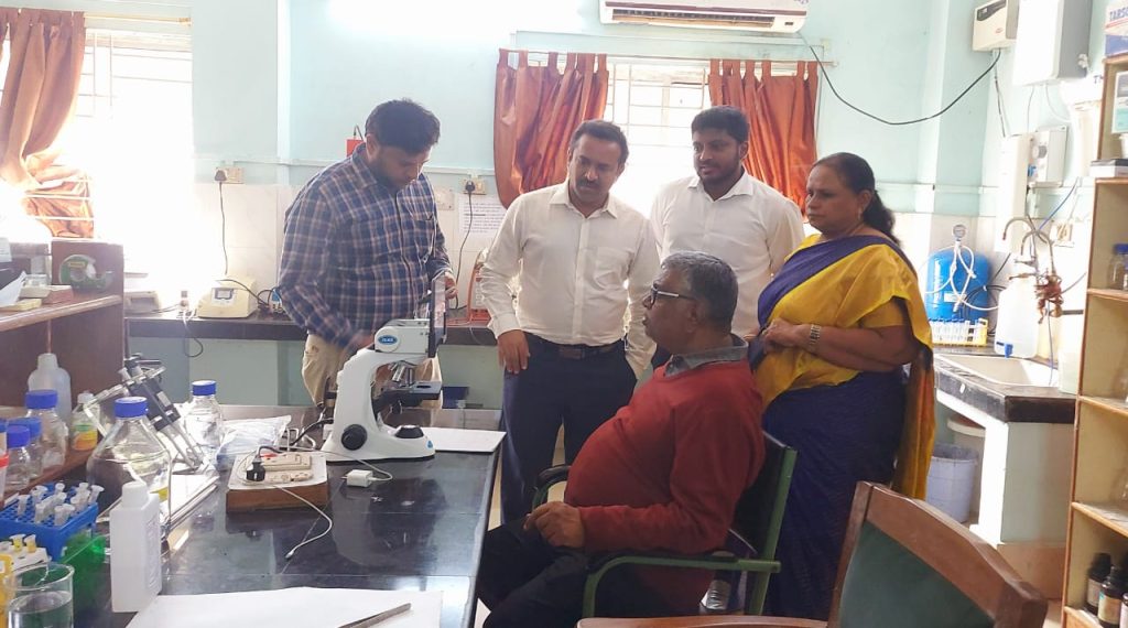
He was on the lookout for a microscope that could fulfil all of his requirements and he came across Cilika. Within no time, Cilika was installed at his workplace, seamlessly integrating into his daily research. What followed was a complete transformation in the way he conducted his research.
How Cilika Transformed Silkworm Screening
From the moment he started using Cilika, Dr. Arun felt the difference. The physical strain he once endured was gone. Instead of bending over an eyepiece for hours, he could now comfortably examine specimens on a large digital display, reducing stress on his neck and back. His eyes, too, felt less fatigued, as the LED screen provided a clearer and more comfortable viewing experience.
The biggest breakthrough, however, was the improvement in efficiency. With Cilika, Dr. Arun could now screen many samples at a stretch, drastically reducing the time spent on each analysis. This efficiency not only benefited his research but also proved invaluable for farmers, who could now screen their own samples and understand the presence of pathogens in their silkworms. With 100% field of view, he no longer had to adjust slides repeatedly to get a complete picture. The high-resolution imaging allowed him to detect abnormalities faster, significantly reducing the time required for screening. The portability of Cilika also meant that he could take it directly to silk farms and conduct screenings on-site, something that was previously impossible with traditional microscopes.
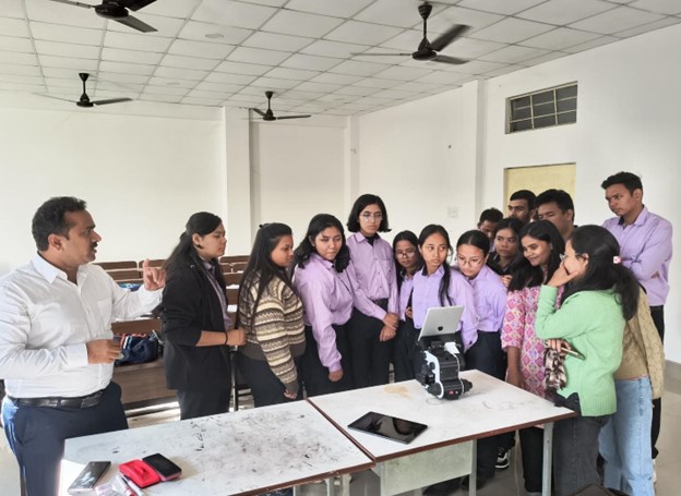
Collaboration also became seamless. The LED screen enabled multiple researchers and students to observe the samples simultaneously, making training sessions more interactive and effective. Moreover, Cilika’s capability as a projection microscope allowed for large-scale demonstrations. The ability to display results on a large screen allowed multiple people to observe the sample at the same time, making it easier to educate farmers about disease prevention and the quality of their silkworm batches. During workshops, instead of students taking turns to peer through a single eyepiece, entire groups could now view and discuss specimens in real time, leading to a more engaging learning experience.
Documentation, another time-intensive task, became easier as well. With Cilika’s digital capabilities, he could capture and annotate images instantly, reducing the manual effort required for record-keeping and analysis.
A New Era for Silkworm Research
With the integration of Cilika microscope, Dr. Arun and his team have experienced remarkable improvements in disease detection. The microscope has not only enhanced routine diagnosis but has also revolutionized on-site diagnosis and training programs for farmers. With greater accuracy, efficiency, and ease of use, Cilika is proving to be a game-changer in every field.
Dr. Arun’s success story is just one example of how Cilika digital microscopes are transforming research across various industries, providing smart, ergonomic, and efficient microscopy solutions.
Why Cilika is the future of digital microscopy?
Cilika is an advanced digital microscope, designed to provide high-resolution imaging, seamless collaboration, and effortless digital integration. Unlike traditional microscopes, Cilika is not just a digital microscope but also a smart microscope, integrating tablet/smartphone compatibility for enhanced functionality.
Featuring 100% field of view (FOV), Cilika ensures that the entire sample can be observed without any loss of image quality. The integration of digital technology eliminates lag, enabling real-time, smooth image viewing.
With an ergonomic design, Cilika significantly reduces physical strain, allowing for prolonged usage without discomfort. One of its type that is cilika portable microscope make it ideal for both laboratory and field settings, enabling routine diagnosis and on-site research. The preloaded Cilika software offers micrometry and annotations, allowing users to analyze and document their findings effortlessly.
Cilika also enhances collaboration and knowledge sharing. Its digital display and projection microscope capabilities allow multiple users to observe samples simultaneously, making it an excellent tool for training, discussions, and real-time group analysis. Whether in research, education, or diagnostics, Cilika ensures that microscopy is no longer limited to a single viewer, fostering better engagement and teamwork.
With its smart design, digital adaptability, and seamless sharing capabilities, Cilika is redefining modern microscopy and empowering professionals across industries.
Beyond its technological advancements, Cilika is making its mark across every state in India, empowering researchers, educators, and pathologists with advanced digital microscopy. Beyond India, we are progressively expanding into the international market, with growing curiosity and interest from the Middle East and Asian continent. As more industries recognize the impact of smart digital microscopy, Cilika continues to push the boundaries of innovation in global research and diagnostics.